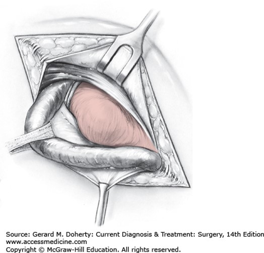
The boundaries of the inguinal canal must be understood to comprehend the principles of hernia repair. In the inguinal canal, the anterior boundary is the external oblique aponeurosis; the posterior boundary is composed of the transversalis fascia with some contribution from the aponeurosis of the transversus abdominis muscle; the inferior border is imparted by the inguinal and lacunar ligaments; and the superior boundary is formed by the arching fibers of the internal oblique musculature.
The internal (or deep) inguinal ring is formed by a normal defect in the transversalis fascia through which the spermatic cord in men and the round ligament in women passes into the abdomen from the extraperitoneal plane. The external (or superficial) ring is inferior and medial to the internal ring and represents an opening of the aponeurosis of the external oblique. The spermatic cord passes from the peritoneum through the internal ring and then caudally into the external ring before entering the scrotum in males.
From superficial to deep, the surgeon first encounters Scarpa's fascia after incising the skin and subcutaneous tissue. Deep to Scarpa's layer is the external oblique aponeurosis, which must be incised and spread to identify the cord structures. The inguinal ligament represents the inferior extension of the external oblique aponeurosis, and extends from the anterior superior iliac spine to the pubic tubercle. The medial extension of the external oblique aponeurosis forms the anterior rectus sheath. The iliohypogastric and ilioinguinal nerves, which provide sensation to the skin, penis, and the upper medial thigh, lie deep to the external oblique aponeurosis in the groin region. The internal oblique aponeurosis is more prominent cephalad in the inguinal canal, and its fibers form the superior border of the canal itself. The cremaster muscle, which envelops the cord structures, originates from the internal oblique musculature. The transversalis abdominis muscle and its fascia represent the true floor of the inguinal canal. Deep to the floor is the preperitoneal space, which houses the inferior epigastric artery and vein, the genitofemoral and lateral femoral cutaneous nerves, and the vas deferens, which traverses this space to join the remaining cord structures at the internal inguinal ring.

Figure legend: Direct inguinal hernia. Inguinal canal opened and spermatic cord retracted inferiorly and laterally to reveal the hernia bulging through the floor of the Hesselbach triangle.
Board Review Questions
1. Poupart’s ligament is composed of fibers from which muscle aponeurosis?
A. Rectus abdominis
B. Transversalis
C. Internal oblique
D. External oblique aponeurosis
2. Which nerve travels with the spermatic cord, entering the inguinal canal at the internal ring, and exiting at the external ring?
A. Iliohypogastric nerve
B. Ilioinguinal nerve
C. Genitofemoral nerve
D. Lateral femoral cutaneous nerve
3. Which of the following is one of the three borders of Hesselbach’s triangle?
A. Superior epigastric artery
B. Edge of the transversalis muscle
C. Inguinal ligament
D. Internal inguinal ring
Answers
1. The correct answer is D. External oblique aponeurosis
2. The correct answer is B. Ilioinguinal nerve
3. The correct answer is C. Inguinal ligament

Create a Free MyAccess Profile
AccessMedicine Network is the place to keep up on new releases for the Access products, get short form didactic content, read up on practice impacting highlights, and watch video featuring authors of your favorite books in medicine. Create a MyAccess profile and follow our contributors to stay informed via email updates.