Like
Be the first to like this
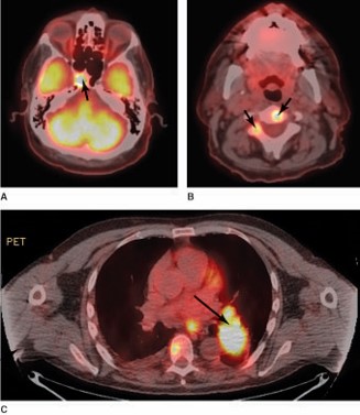
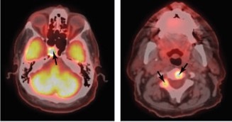
A 62-year-old man presented with a progressive right abduction deficit. Magnetic resonance imaging (MRI) showed a right cavernous sinus mass, suspected to be metastatic. Positron emission tomography-computed tomography (PET-CT) revealed the right cavernous sinus lesion and multiple metastases (short arrows), with a lung mass presumed to be the primary neoplasm (long arrow), shown in these images through the A) and B) brain and B) and C) chest. Note the normal high metabolic activity of the A) brain.
Source: Martin TJ, Corbett JJ. Practical Neuroophthalmology; 2013.


Create a Free MyAccess Profile
AccessMedicine Network is the place to keep up on new releases for the Access products, get short form didactic content, read up on practice impacting highlights, and watch video featuring authors of your favorite books in medicine. Create a MyAccess profile and follow our contributors to stay informed via email updates.