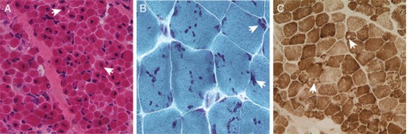
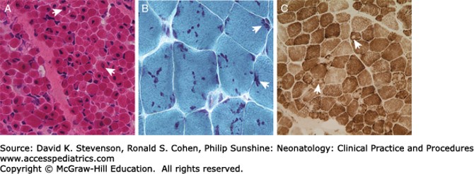
X-linked myotubular myopathy, nemaline myopathy, and multiminicore myopathy. Representative light microscopic images from muscle biopsies obtained during infancy and early childhood from patients with various forms of congenital myopathy. A, A hematoxylin and eosin stain of a muscle biopsy from a child with X-linked myotubular myopathy. The histology is characterized by an abundance of centralized nuclei, examples of which are indicated by the arrows. B, A modified Gomori trichrome stain illustrates the abundance of nemaline rods (arrows) that are typically found in children with nemaline myopathy. C, Multiple minicores (arrows) are seen on cytochrome oxidase staining of muscle tissue from a child with multiminicore myopathy.
Source: Stevenson DK, Cohen RS, Sunshine P. Neonatology: Clinical Practice and Procedures; 2015.


Create a Free MyAccess Profile
AccessMedicine Network is the place to keep up on new releases for the Access products, get short form didactic content, read up on practice impacting highlights, and watch video featuring authors of your favorite books in medicine. Create a MyAccess profile and follow our contributors to stay informed via email updates.