Coronary Artery Disease (CAD) on PET and CT

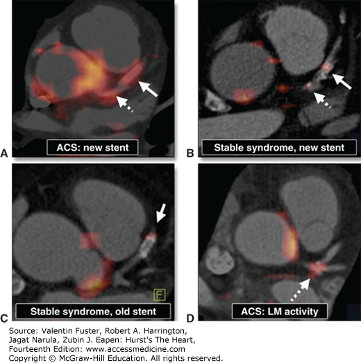
FIGURE 19–65.
18F-fluorodeoxyglucose (FDG) positron emission tomography (PET)/computed tomography (CT) images of coronary arterial 18F-FDG uptake. Intense 18F-FDG uptake is seen in the left main coronary artery and the stented culprit lesion in a patient with acute coronary syndrome (ACS) (A). Uptake is less intense in another patient with stable coronary artery disease and a recent stent placement (B), whereas only mild uptake is seen in a patient with coronary artery disease and an old stent (C). Mild 18F-FDG uptake is noted in the trifurcation of the left main (LM) coronary artery in a patient with ACS (D). Reproduced with permission from Rogers IS, Nasir K, Figueroa AL, et al: Feasibility of FDG imaging of the coronary arteries: comparison between acute coronary syndrome and stable angina. JACC Cardiovasc Imaging. 2010 Apr;3(4):388-397.236

Read More about Imaging Studies to Diagnose CAD in Context:
Hurst’s the Heart, 14e: Chapter 19. Positron Emission Tomography in Heart Disease
Read about Imaging Studies to Diagnose CAD:
Harrison’s Principles of Internal Medicine, 20e: Chapter 236. Noninvasive Cardiac Imaging: Echocardiography, Nuclear Cardiology, and Magnetic Resonance/ Computed Tomography Imaging
Principles and Practice of Hospital Medicine, 2e: Chapter 115. Advanced Cardiothoracic Imaging


Create a Free MyAccess Profile
AccessMedicine Network is the place to keep up on new releases for the Access products, get short form didactic content, read up on practice impacting highlights, and watch video featuring authors of your favorite books in medicine. Create a MyAccess profile and follow our contributors to stay informed via email updates.