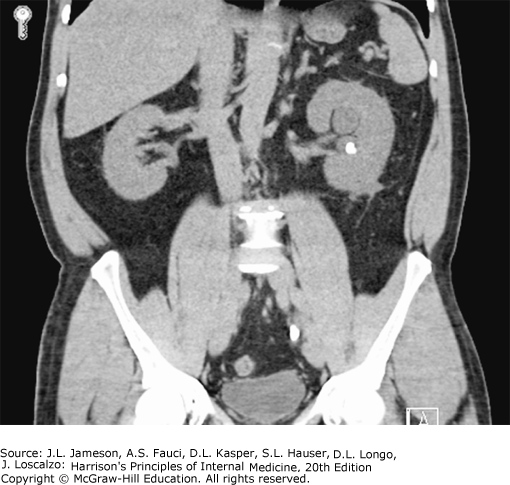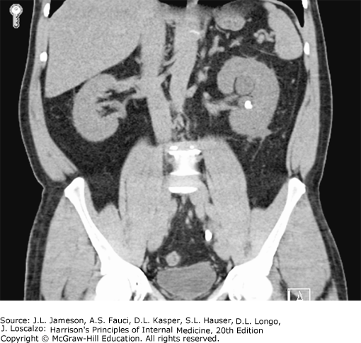
Coronal noncontrast CT image from a patient who presented with left-sided renal colic. An obstructing calculus, present in the distal left ureter at the level of S1, measures 10 mm in maximal dimension. There is severe left hydroureteronephrosis and associated left perinephric fat stranding. In addition, there is a nonobstructing 6-mm left renal calculus in the interpolar region. (Image courtesy of Dr. Stuart Silverman, Brigham and Women’s Hospital.)

Read more about renal calculi in context:
Harrison’s Principles of Internal Medicine, 20e: Chapter 312. Nephrolithiasis
Read more about renal calculi:
The Color Atlas and Synopsis of Family Medicine, 3e: Chapter 70. Kidney Stones
Current Medical Diagnosis and Treatment 2020: Chapter 23-05. Urinary Stone Disease


Create a Free MyAccess Profile
AccessMedicine Network is the place to keep up on new releases for the Access products, get short form didactic content, read up on practice impacting highlights, and watch video featuring authors of your favorite books in medicine. Create a MyAccess profile and follow our contributors to stay informed via email updates.