Adenosine Stress/Rest Myocardial Perfusion Single-Photon Emission Computed Tomography

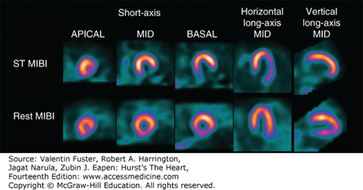
Case example illustrating value of stress testing ST when rest myocardial perfusion imaging is normal in the acute chest pain patient. Shown are adenosine stress/rest myocardial perfusion single-photon emission computed tomography images in a 55-year-old man with multiple risk factors for coronary artery disease presenting to the emergency department with 2 days of chest discomfort at rest. Rest sestamibi (MIBI) demonstrated minimal (borderline) perfusion defect in the inferior wall. Stress imaging revealed evidence of severe and extensive ischemia in the inferior and inferolateral wall (41% of the left ventricle). Coronary angiography revealed an occluded right coronary artery that was successfully stented.
Read about myocardial imaging studies in context: Hurst’s the Heart, 14e: Chapter 18. Nuclear Cardiology
Read more about myocardial imaging studies:
Harrison’s Principles of Internal Medicine, 20e: Chapter 236: Noninvasive Cardiac Imaging: Echocardiography, Nuclear Cardiology, and Magnetic Resonance/Computed Tomography Imaging
Introduction to Diagnostic Radiology: Chapter 2. Introduction to Nuclear Medicine
Melanie Allison is the Executive Manager of Education & Learning with McGraw Hill. She earned her Doctor of Nursing Practice (DNP) degree and Post-Master’s Certificate in Nursing Education from The Johns Hopkins University. She earned her Master of Science in Nursing (MSN) degree, specializing as an acute care nurse practitioner (ACNP), from Vanderbilt University. Melanie has more than 20 years of experience as a registered nurse and nurse practitioner in adult cardiology and advanced lipid management. She is a part-time faculty member at a top school of nursing where she has taught for more than 16 years.


Create a Free MyAccess Profile
AccessMedicine Network is the place to keep up on new releases for the Access products, get short form didactic content, read up on practice impacting highlights, and watch video featuring authors of your favorite books in medicine. Create a MyAccess profile and follow our contributors to stay informed via email updates.