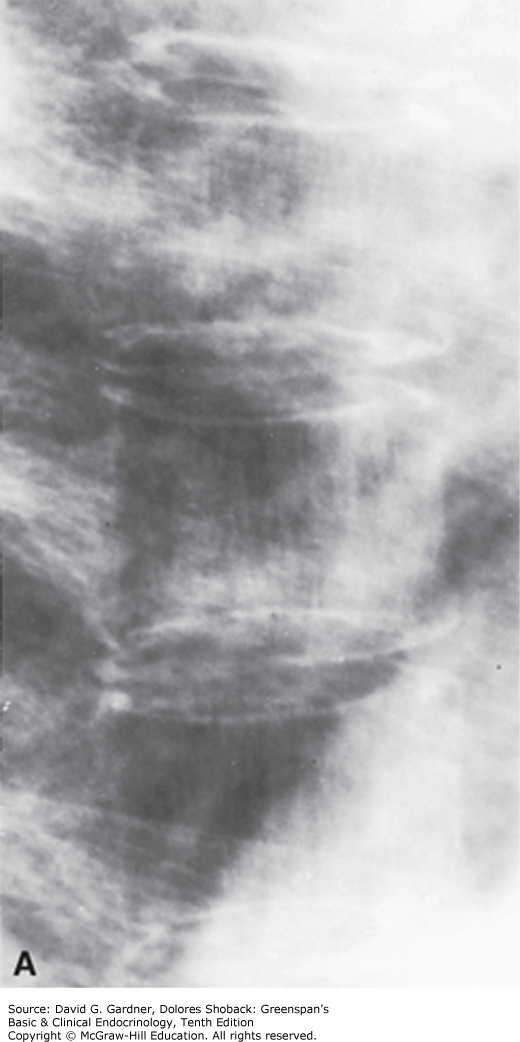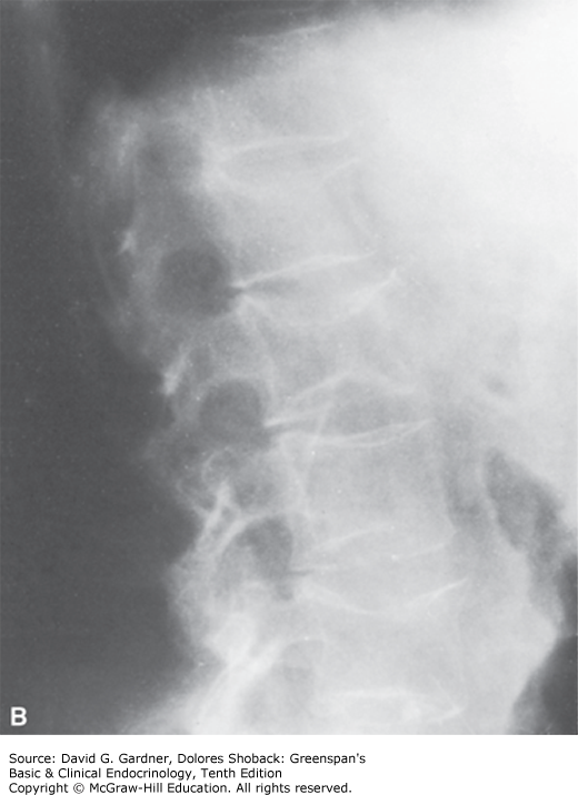
A. Magnified x-rays of thoracic vertebrae from a woman with osteoporosis. Note the relative prominence of vertical trabeculae and the absence of horizontal trabeculae. B. Lateral x-ray of the lumbar spine of a woman with postmenopausal osteoporosis. Note the increased density of the superior and inferior cortical margins of vertebrae, the marked demineralization of vertebral bodies, and the central compression of articular surfaces of vertebral bodies by intervertebral disks. (Used with permission from Dr. G. Gordan.)


Read more about osteoporosis in context:
Greenspan’s Basic and Clinical Endocrinology, 10e: Chapter 8. Metabolic Bone Disease
Read more about osteoporosis:
Hazzard’s Geriatric Medicine and Gerontology, 7e: Chapter 118. Osteoporosis
Williams Gynecology, 3e: Chapter 21. Menopausal Transition
Harrison’s Principles of Internal Medicine, 20e: Chapter 404. Osteoporosis


Create a Free MyAccess Profile
AccessMedicine Network is the place to keep up on new releases for the Access products, get short form didactic content, read up on practice impacting highlights, and watch video featuring authors of your favorite books in medicine. Create a MyAccess profile and follow our contributors to stay informed via email updates.