Brain MRI Findings in Patients with Dementia

Top Image: Coronal T1-weighted MRI of a 74-year-old man with moderate Alzheimer-type dementia. Diffuse cerebral and hippocampal atrophy with ex vacuo ventricular and cortical sulcal dilation is noted.
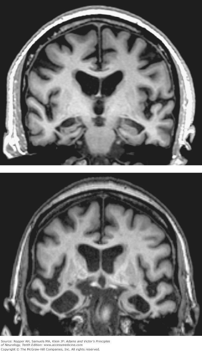
Bottom Image: Coronal T1-weighted MRI of a 70-year-old woman with behavioral variant frontotemporal lobar dementia. Atrophy of the right greater than left temporal lobes is out of proportion to atrophy of the frontal and parietal lobes.
Click this link to read more about dementia in context: Adam and Victor's Principles of Neurology, 10e> Chapter 39: Degenerative Diseases of the Nervous System
Click this link to read more about dementia: Hazzard's Geriatric Medicine and Gerontology, 7e > Chapter 66: Dementia Including Alzheimer Disease
Melanie Allison is the Executive Manager of Education & Learning with McGraw Hill. She earned her Doctor of Nursing Practice (DNP) degree and Post-Master’s Certificate in Nursing Education from The Johns Hopkins University. She earned her Master of Science in Nursing (MSN) degree, specializing as an acute care nurse practitioner (ACNP), from Vanderbilt University. Melanie has more than 20 years of experience as a registered nurse and nurse practitioner in adult cardiology and advanced lipid management. She is a part-time faculty member at a top school of nursing where she has taught for more than 16 years.


Create a Free MyAccess Profile
AccessMedicine Network is the place to keep up on new releases for the Access products, get short form didactic content, read up on practice impacting highlights, and watch video featuring authors of your favorite books in medicine. Create a MyAccess profile and follow our contributors to stay informed via email updates.