Dermatology Question of the Week: Pediatric Problems

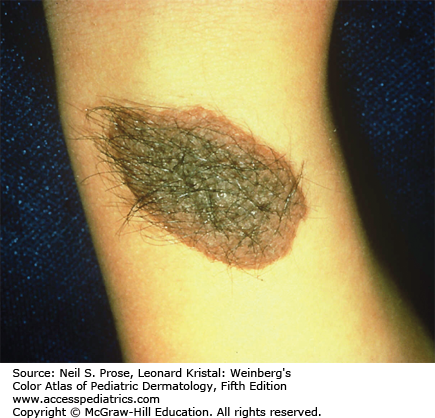
You are consulted to evaluate a newborn who has several 2-5cm brown plaques with hair similar to the picture below. The primary team and family are wondering if there is anything to do for these spots.

Which of the following is the best initial step?
A. Reassurance and monitoring for change
B. Skin biopsy
C. MRI brain and spine without contrast
D. Echocardiogram
Rationale: The description and photograph are consistent with congenital nevi. Having giant/large congenital nevi with satellitosis OR having multiple medium-sized congenital nevi increases the risk of patients having and developing neurocutaneous melanosis (NCM). The best screening test for this is an MRI of the brain and spine; common MRI findings of NCM include intraparenchymal melanosis and leptomeningeal enhancement. Early screening can be helpful and avoid the use of contrast or sedation; contrast is required to visualize melanin after myelinization which occurs around 6 months of age.
Correct answer: C MRI brain and spine without contrast
As noted above, early screening with an MRI is appropriate for evaluating NCM. Patients may develop hydrocephalus, seizures, brain malformations such as Dandy-Walker malformation, or even melanoma. Neurosurgical involvement early on is critical to address brain abnormalities and assess for signs of intracranial pressure as the mortality rate for patients who develop symptoms is high.
Incorrect answers:
A. Although reassurance is typically appropriate when patients have a single congenital melanocytic nevus, this patient has multiple congenital melanocytic nevi which can be associated with NCM.
B. Biopsy of any changing congenital nevus would be appropriate. A biopsy of the melanocytic proliferation in the brain/spine could be considered if the patient is undergoing a neurological procedure and if the area is easy to access. Biopsy of any new melanocytic nodule or proliferation in the brain/spine may be appropriate as well to rule out melanoma.
D. Carney complex is an autosomal dominant condition where patients develop multiple lentigines/blue nevi, cutaneous and extracutaneous myxomas, thyroid and other endocrine tumors. Screening in this condition for cardiac myxomas with echocardiogram is appropriate however this condition does not fit our clinical vignette.
Additional reading at Fitzpatrick's Dermatology Chapter 115: Melanocytic Nevi
References:
1. Mologousis MA, Tsai SY-C, Tissera KA, Levin YS, Hawryluk EB. Updates in the Management of Congenital Melanocytic Nevi. Children. 2024; 11(1):62. https://doi.org/10.3390/children11010062
2. Jahnke MN, et al. Care of Congenital Melanocytic Nevi in Newborns and Infants: Review and Management Recommendations. Pediatrics. 2021 Dec 1;148(6):e2021051536. doi: 10.1542/peds.2021-051536.
3. Rahman RK, Majmundar N, Ghani H, San A, Koirala M, Gajjar AA, Pappert A, Mazzola CA. Neurosurgical management of patients with neurocutaneous melanosis: a systematic review. Neurosurg Focus. 2022 May;52(5):E8. doi: 10.3171/2022.2.FOCUS21791. PMID: 35535823.
4. Habibi Z, Ebrahimi H, Meybodi KT, Yaghmaei B, Nejat F. Clinical Follow-Up of Patients with Neurocutaneous Melanosis in a Tertiary Center; Proposed Modification in Diagnostic Criteria. World Neurosurg. 2021 Feb;146:e1063-e1070. doi: 10.1016/j.wneu.2020.11.091. Epub 2020 Nov 24. PMID: 33246180.

Create a Free MyAccess Profile
AccessMedicine Network is the place to keep up on new releases for the Access products, get short form didactic content, read up on practice impacting highlights, and watch video featuring authors of your favorite books in medicine. Create a MyAccess profile and follow our contributors to stay informed via email updates.