Dermatology Question of the Week: Medical Mysteries

A 45-year-old male presents with a three-month history of painful oral ulcers as shown below.
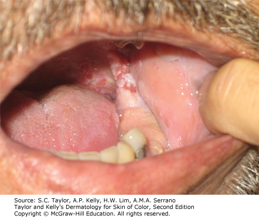
He occasionally gets similar lesions on his gingiva and lips and has noted some blisters that rupture easily on his trunk. A biopsy from an intact blister shows intraepidermal acantholysis and direct immunofluorescence reveals intercellular IgG deposits in the epidermis. What is the most likely diagnosis?
A. Bullous pemphigoid
B. Pemphigus vulgaris
C. Pemphigus foliaceus
D. Lichen planus
E. Linear IgA bullous dermatosis
F. Epidermolysis bullosa acquisita
Rationale:
This question tests the ability to distinguish between autoimmune blistering diseases and inflammatory dermatoses based on clinical, histologic, and immunofluorescence findings. There are various pathogenic causes of oral ulcerations including pemphigus vulgaris, lichen planus, bullous pemphigoid, as well as fungal and viral infections. The importance of DIF cannot be underscored as many of these diagnoses can appear similarly on H&E. At times, serum antibody studies or indirect immunofluorescence may be helpful.
Correct answer:
B. Pemphigus vulgaris is characterized by flaccid blisters, mucosal erosions, and a positive Nikolsky sign. Histologically, it shows intraepidermal acantholysis, and direct immunofluorescence demonstrates intercellular IgG deposits in a "chicken wire" pattern. Oral involvement is a hallmark feature of pemphigus vulgaris.
Incorrect answers:
A. Bullous pemphigoid typically presents with tense blisters rather than flaccid blisters, and mucosal involvement is less common. Direct immunofluorescence shows linear IgG and C3 deposits along the basement membrane zone. Bullous pemphigoid is typically seen in elderly patients although cases in younger individuals have been reported.
C. Pemphigus foliaceus presents with superficial flaccid blisters and erosions typically located on seborrheic areas such as the face, scalp, and chest. Notably, pemphigus foliaceous rarely involves mucosa. Direct immunofluorescence shows intercellular IgG deposits similar to pemphigus vulgaris.
D. Cutaneous lichen planus presents with violaceous, flat-topped papules and plaques, commonly involving the wrists and shins. Oral involvement in the erosive variant of lichen planus can appear similarly to pemphigus. Histologically, it shows a band-like lymphocytic infiltrate at the dermo-epidermal junction. Direct immunofluorescence reveals fibrin or shaggy deposits of fibrinogen at the basement membrane zone, not intercellular IgG deposits.
E. Linear IgA bullous dermatosis typically presents with annular or grouped vesicles and bullae, often on extensor surfaces. Direct immunofluorescence shows linear IgA deposition along the basement membrane zone.
F. Epidermolysis bullosa acquisita presents with tense blisters in trauma-prone areas. Direct immunofluorescence reveals linear IgG and/or C3 deposits at the dermoepidermal junction.
Additional reading at Fitzpatrick's Dermatology Chapter 52: Pemphigus and Dermatology Skin of Color Chapter 32: Acquired Bullous Diseases
I am a general dermatologist practicing at an academic medical center in Danville, PA. I see both adult and pediatric patients and also perform various surgical procedures. I have a special interest in Hidradenitis Suppurativa (HS) and am the founder of our HS specialty clinic where we treat patients medically and surgically (deroofing, excision, staged CO2 excision with marsupialization, Nd:YAG). I am actively involved in resident education and am passionate about medical education. Additional interests include autoimmune bullous disorders, dermatomyositis, and procedural dermatology.

Create a Free MyAccess Profile
AccessMedicine Network is the place to keep up on new releases for the Access products, get short form didactic content, read up on practice impacting highlights, and watch video featuring authors of your favorite books in medicine. Create a MyAccess profile and follow our contributors to stay informed via email updates.