Like
Be the first to like this
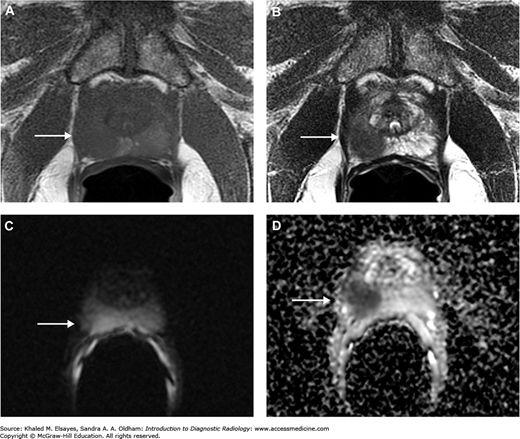
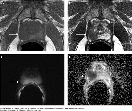
MRI with endorectal coil of a patient with prostate cancer. (A) Axial T1WI. (B) Axial T2WI. (C) DWI. (D) ADC map reveals a well-defined focal lesion exhibiting an isointense signal on T1WI and hypointense on T2WI, with restricted diffusion (arrow).
Read more about prostate cancer in context:
Introduction to Diagnostic Radiology: Chapter 6. Genitourinary System
Read more about prostate cancer:
Current Medical Diagnosis and Treatment 2020: Chapter 39-17. Prostate Cancer
Current Diagnosis and Treatment: Geriatrics, 2e: Chapter 40. Benign Prostatic Hyperplasia and Prostate Cancer
Harrison’s Principles of Internal Medicine, 20e: Chapter 83. Benign and Malignant Diseases of the Prostate


Create a Free MyAccess Profile
AccessMedicine Network is the place to keep up on new releases for the Access products, get short form didactic content, read up on practice impacting highlights, and watch video featuring authors of your favorite books in medicine. Create a MyAccess profile and follow our contributors to stay informed via email updates.