Muscle and Nerve Histopathology Diagnosis
What's your diagnosis?

Like
Be the first to like this

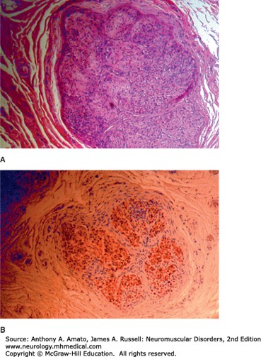
Neurofibroma. The nerve fascicle has a lobulated appearance, H&E stain (A). The cells have wavy, elongated nuclei, and the background material is loosely arranged and myxoid. Bands of thick collagen are apparent in the center of the tumor. Some of the proliferating tumor cells are immunoreactive for S-100, suggesting Schwann cell origin (B).
Source: Amato AA, Russell JA. Neuromuscular Disorders, 2e; 2015.


Create a Free MyAccess Profile
AccessMedicine Network is the place to keep up on new releases for the Access products, get short form didactic content, read up on practice impacting highlights, and watch video featuring authors of your favorite books in medicine. Create a MyAccess profile and follow our contributors to stay informed via email updates.