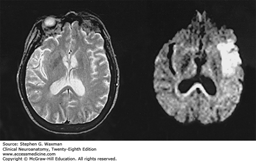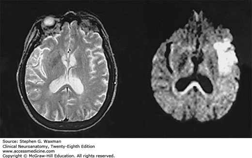

Cerebral infarct shown by diffusion-weighted imaging (DWI). On the left, a conventional MRI (T2-weighted image) 3 hours after stroke onset shows no lesions. On the right, DWI 3 hours after stroke onset shows extensive hyperintensity indicative of acute ischemic injury. (Reproduced, with permission, from Warach S, et al: Acute human stroke studied by whole brain echo planar diffusion weighted MRI. Ann Neurol 1995;37:231.)
Read about stroke in context:
Clinical Neuroanatomy, 28e: Chapter 22. Imaging of the Brain
Read about stroke:
Harrison’s Principles of Internal Medicine, 20e: Chapter 420. Ischemic Stroke
Principles and Practice of Hospital Medicine, 2e: Chapter 209. Transient Ischemic Attack and Stroke
Tintinalli’s Emergency Medicine: A Comprehensive Study Guide, 8e: Chapter 167. Stroke Syndromes


Create a Free MyAccess Profile
AccessMedicine Network is the place to keep up on new releases for the Access products, get short form didactic content, read up on practice impacting highlights, and watch video featuring authors of your favorite books in medicine. Create a MyAccess profile and follow our contributors to stay informed via email updates.