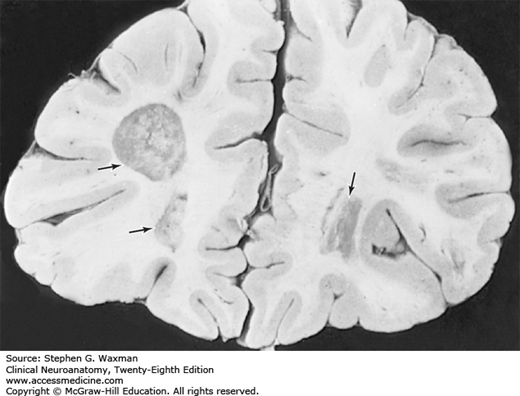
Brain Imaging of Demyelination in Multiple Sclerosis

Areas of demyelination of the white matter (arrows) in the frontal lobe of a 54-year-old man with multiple sclerosis.
Read more about multiple sclerosis in context:
Clinical Neuroanatomy, 28e: Chapter 25. Discussion of Cases
Read more about multiple sclerosis:
Harrison’s Principles of Internal Medicine, 20e: Chapter 436. Multiple Sclerosis
Principles and Practice of Hospital Medicine, 2e: Chapter 212. Multiple Sclerosis
Adams and Victor's Principles of Neurology, 10e: Chapter 36. Multiple Sclerosis and Other Inflammatory Demyelinating Diseases
Melanie Allison is the Executive Manager of Education & Learning with McGraw Hill. She earned her Doctor of Nursing Practice (DNP) degree and Post-Master’s Certificate in Nursing Education from The Johns Hopkins University. She earned her Master of Science in Nursing (MSN) degree, specializing as an acute care nurse practitioner (ACNP), from Vanderbilt University. Melanie has more than 20 years of experience as a registered nurse and nurse practitioner in adult cardiology and advanced lipid management. She is a part-time faculty member at a top school of nursing where she has taught for more than 16 years.


Create a Free MyAccess Profile
AccessMedicine Network is the place to keep up on new releases for the Access products, get short form didactic content, read up on practice impacting highlights, and watch video featuring authors of your favorite books in medicine. Create a MyAccess profile and follow our contributors to stay informed via email updates.