Week 4 Image
What’s your diagnosis?

Like
Be the first to like this
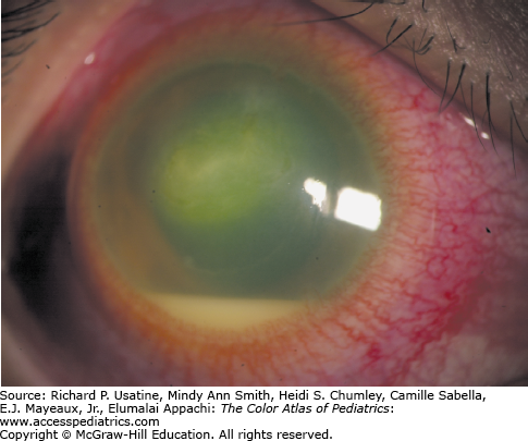
Answer: Hypopyon with severe anterior uveitis, showing layering of leukocytes and fibrinous debris in the anterior chamber. May be sterile or infectious. An intense ciliary flush is seen. Most commonly seen in HLA-B27-positive patients with uveitis. Hypopyon may also be a presenting sign of malignancy (retinoblastoma and lymphoma).
From: Usatine RP, Sabella C, Smith M, Mayeaux EJ, Jr., Chumley HS, Appachi E. The Color Atlas of Pediatrics; 2015 Available at: http://accesspediatrics.mhmedical.com/ViewLarge.aspx?figid=94707656 Accessed: February 13, 2018


Create a Free MyAccess Profile
AccessMedicine Network is the place to keep up on new releases for the Access products, get short form didactic content, read up on practice impacting highlights, and watch video featuring authors of your favorite books in medicine. Create a MyAccess profile and follow our contributors to stay informed via email updates.