Coccydynia


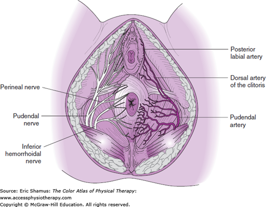
Scenario: A 25-year-old woman delivered her first baby vaginally. She had epidural anesthesia during the delivery. During the delivery, she heard a loud “pop” noise. After the epidural wore off, she felt a severe pain in her rear end. She was unable to sit on the edge of the hospital bed and had severe pain when she attempted to sit in a chair.
Question: What is the most common type of imaging used to diagnose coccydynia?
A. Dynamic Radiograph
B. MRI
C. Static Radiograph
D. CT scan
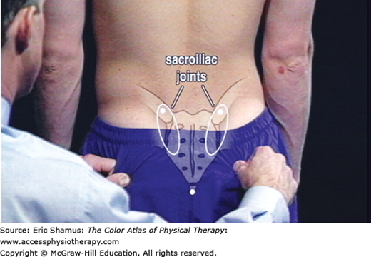
Answer with rationale: A. Dynamic radiograph. Most often of the coccyx position in sitting versus standing. Dynamic radiograph is chosen because it helps the clinician to visualize abnormal mobility. This imaging type also highlights the presence of any bone spurs that may occur.
For more information see Chapter 119: Coccydynia in The Color Atlas of Physical Therapy

Create a Free MyAccess Profile
AccessMedicine Network is the place to keep up on new releases for the Access products, get short form didactic content, read up on practice impacting highlights, and watch video featuring authors of your favorite books in medicine. Create a MyAccess profile and follow our contributors to stay informed via email updates.